| Deletions are marked like this. | Additions are marked like this. |
| Line 210: | Line 210: |
|
- - - |
|
| Line 248: | Line 251: |
|
- - - |
|
| Line 279: | Line 285: |
|
- - - |
|
| Line 285: | Line 294: |
|
- - - |
|
| Line 287: | Line 299: |
|
This section will run you through using the stats directory in the subjects to perform group stats of certain structures that may be of interest to your study. The following commands will help you combine the data of the subjects you are analyzing into one table that will be easily read into a spreadsheet program. We have considered 6 subjects as examples (004, 021, 040, 067, 080, 092) in the following sections. Set your SUBJECTS_DIR to the path where you have your subjects to be analyzed. '' '' ---- '' '' '' '' {{{ }}} ---- '' '' '' '' |
This section will run you through using the stats directory of the subjects to perform group stats of certain structures that may be of interest to your study. The following commands will help you combine the data of the subjects you are analyzing into one table that will be easily read into a spreadsheet program. We have considered 6 subjects as examples (004, 021, 040, 067, 080, 092) in the following sections. Set your SUBJECTS_DIR to the path where you have your subjects to be analyzed. ---- {{{ setenv SUBJECTS_DIR $TUTORIAL_DATA/buckner_data/tutorial_subjs/group_analysis_tutorial cd $SUBJECTS_DIR }}} ---- |
| Line 301: | Line 313: |
|
'' '' '' '' {{{ }}} ---- '' '' where 11, 17 and 18 correspond to the segmentation label of left caudate, left hippocampus and left amygdala respectively. (You can create a table with all of the labels, not just these three, by omitting the --segno part.) Click [[FsTutorial/AnatomicalROI/FreeSurferColorLUT|here]] if you would like to view the list of labels and their corresponding ID numbers again. The file {{{aseg.vol.table}}} is your output - a text file consisting of the subjects mentioned in the command above and the values for the structures requested. The information in this text file is formatted so it can be easily imported into a spreadsheet program (often used as input for many statistical analysis programs). If you do the {{{ls}}} command, you should see that the text file {{{aseg.vol.table}}} has been created. To see what the file looks like, do: '' '' ---- '' '' '' '' {{{ }}} ---- '' '' To to load the resulting table into a spreadsheet, run: '' '' ---- '' '' '' '' {{{ }}} ---- '' '' '''Note:''' the ''gnumeric'' command is meant to be run on a Linux machine. Mac users could install !OpenOffice 3.0 to open the table in a spreadsheet and run the command '' '' ---- '' '' '' '' {{{ }}} ---- '' '' In the table, the first cell is ''volume'' indicating that the measure is a volume in mm3. In addition to segmentations, you can also get IntraCranialVol (ICV) and BrainSegVol into the table. About the subject IDs, you'll notice that in the examples we've considered here, each subject is a 3 digit number. Therefore ''gnumeric'' thinks it is a number and removes leading 0s. This is a gnumeric issue but probably it would not happen if subject names had characters in them instead of integers. '' '' |
{{{ asegstats2table --subjects 004 021 040 067 080 092 \ --segno 11 17 18 \ --tablefile aseg.vol.table }}} ---- where 11, 17 and 18 correspond to the segmentation label of left caudate, left hippocampus and left amygdala respectively. (You can create a table with all of the labels, not just these three, by omitting the --segno part.) Click [[FsTutorial/AnatomicalROI/FreeSurferColorLUT|here]] if you would like to view the list of labels and their corresponding ID numbers again. The file {{{aseg.vol.table}}} is your output - a text file consisting of the subjects mentioned in the command above and the values for the structures requested along with the measures in the header (i.e. BrainSegVol). The information in this text file is formatted so it can be easily imported into a spreadsheet program (often used as input for many statistical analysis programs). If you do the {{{ls}}} command, you should see that the text file {{{aseg.vol.table}}} has been created. To see what the file looks like, do: ---- {{{ less aseg.vol.table }}} ---- To load the resulting table into a spreadsheet, run: ---- {{{ gnumeric aseg.vol.table }}} ---- '''Note:''' the ''gnumeric'' command is meant to be run on a Linux machine. Mac users could install !OpenOffice 3.0 to open the table in a spreadsheet and run the command. ---- {{{ /Applications/OpenOffice.org.app/Contents/MacOS/scalc aseg.vol.table }}} ---- In the table, the first cell is ''volume'' indicating that the measure is a volume in mm3. About the subject IDs, you'll notice that in the examples we've considered here, each subject is a 3 digit number. Therefore ''gnumeric'' thinks it is a number and removes leading 0s. This is a gnumeric issue but probably it would not happen if subject names had characters in them instead of integers. |
| Line 333: | Line 345: |
|
Purpose of this section is to demonstrate how you can change what measure you collect for your spreadsheet from volume to mean intensity using the ''asegstats2table'' command. '' '' ---- '' '' '' '' {{{ }}} ---- '' '' Things to do: '' '' 1. You can load the table into a spreadsheet as explained in the previous section. '' '' 1. Refer to the file ''FreeSurferColorLUT.txt'' for the segmentation labels and the corresponding subcortical structures. '' '' ---- '' '' '' '' {{{ }}} ---- '' '' For tutorial purposes, click [[FsTutorial/AnatomicalROI/FreeSurferColorLUT|here]] to view the contents of the file. '' '' |
The purpose of this section is to demonstrate how you can change what measure you collect for your spreadsheet from volume to mean intensity using the ''asegstats2table'' command. ---- {{{ asegstats2table \ --subjects 004 021 040 067 080 092 \ --segno 11 17 18 \ --meas mean \ --tablefile aseg.mean-intensity.table }}} ---- You can load the table into a spreadsheet as explained in the previous section or view aseg.mean-intensity.table with a text editor, like gedit. |
| Line 358: | Line 363: |
|
'' '' '' '' {{{ }}} ---- '' '' Things to do: '' '' 1. You can load the table into a spreadsheet. '' '' 1. Refer to the file ''FreeSurferColorLUT.txt'' for the segmentation labels and the corresponding subcortical structures. '' '' ---- '' '' '' '' {{{ }}} ---- '' '' For tutorial purposes, click [[FsTutorial/AnatomicalROI/FreeSurferColorLUT|here]] to view the contents of the file '' '' |
{{{ asegstats2table \ --subjects 004 021 040 067 080 092 \ --segno 3007 3021 3022 4022 \ --stats wmparc.stats \ --tablefile wmparc.vol.table }}} ---- This prints out stats on the white matter parcellation. |
| Line 377: | Line 374: |
|
This section explains how to create a table of the surface area of each cortical parcellation in the Desikan atlas (surface area is the default measure). '' '' ---- '' '' '' '' {{{ }}} ---- '' '' You can now load the resulting table into a spreadsheet. '' '' |
This section demonstrates how to create a table of the surface area of each cortical parcellation in the Desikan atlas (surface area is the default measure). ---- {{{ aparcstats2table --hemi lh \ --subjects 004 021 040 067 080 092 \ --tablefile lh.aparc.area.table }}} ---- Feel free to take a look at those results. |
| Line 388: | Line 386: |
|
The purpose of this section is to show how to change the summary measure and the parcellation atlas. '' '' ---- '' '' '' '' {{{ }}} ---- '' '' You can now load the resulting table into a spreadsheet. '' '' |
The purpose of this section is to show how to change the summary measure (in this case, to thickness) and the parcellation atlas (to Destrieux's atlas). ---- {{{ aparcstats2table --hemi lh \ --subjects 004 021 040 067 080 092 \ --meas thickness \ --parc aparc.a2009s \ --tablefile lh.aparc.a2009.thickness.table }}} ---- You can now load the resulting table into a spreadsheet. |
1. Anatomical ROI analysis
This tutorial gives a brief introduction to anatomical ROI analysis which includes working with label files, extracting ROI measures from the anatomical data, group statistics etc.
1.1. Preparations
1.1.1. If You're at an Organized Course
If you are taking one of the formally organized courses, everything has been set up for you on the provided laptop. The only thing you will need to do is run the following commands in every new terminal window (aka shell) you open throughout this tutorial. Copy and paste the commands below to get started:
setenv SUBJECTS_DIR $TUTORIAL_DATA/buckner_data/tutorial_subjs/group_analysis_tutorial
cd $SUBJECTS_DIR
To copy: Highlight the command in the box above, right click and select copy (or use keyboard shortcut Ctrl+c), then use the middle button of your mouse to click inside the terminal window (this will paste the command). Press enter to run the command. These two commands set the SUBJECTS_DIR variable to the directory where the data is stored and then navigates into this directory. You can now skip ahead to the tutorial (below the gray line).
1.1.2. If You're not at an Organized Course
If you are NOT taking one of the formally organized courses, then to follow this exercise exactly be sure you've downloaded the tutorial data set before you begin. If you choose not to download the data set you can follow these instructions on your own data, but you will have to substitute your own specific paths and subject names. These are the commands that you need to run before getting started:
tcsh
source your_freesurfer_dir/SetUpFreeSurfer.csh
setenv SUBJECTS_DIR $TUTORIAL_DATA/buckner_data/tutorial_subjs/group_analysis_tutorial
cd $SUBJECTS_DIR
Notice the command to open tcsh. If you are already running the tcsh command shell, then the 'tcsh' command is not necessary. If you are not using the tutorial data you should set your SUBJECTS_DIR to the directory in which the recon(s) of the subject(s) you will use for this tutorial are located.
1.2. Relationship between segmentation, parcellation and LookUp Table (LUT)
In this exercise, you will examine a segmentation, parcellation, and color lookup table to understand how they are related. Open the subject in freeview using the following command:
freeview -v 004/mri/orig.mgz \
004/mri/aparc+aseg.mgz:colormap=lut:opacity=0.4 \
-f 004/surf/lh.white:annot=aparc.annot
Finally, run the following command to display the contents of LUT (in another new terminal window)
less $FREESURFER_HOME/FreeSurferColorLUT.txt
You can hit the 'Page Up' and 'Page Down' buttons to see the rest of the file. Or click here to view the contents of the file. (To exit the less command, hit the 'q' button.)
Things to do -- Navigating between freeview and LUT:<<BR>>
- Choose the coronal view and click on a cortical structure in the brain.
- See the structure name next to 'aparc+aseg' in the Cursor section below the main viewing window. For example, it may say ctx-lh-precentral. Notice which hemisphere is specified.
- Look at the number listed immediately after the 'aparc+aseg'. For example, it may say 1024.
- Find that value in the LUT, which you have opened using the command mentioned above.
- Verify that it is the same structure you chose in freeview.
- Do the same with a subcortical structure of your choice.
You can close freeview once you are done. To get out of the less command, type 'q' for quit.
1.3. Label files
This section gives the details of commands to load an existing label file in text editor and freeview. To map a label, such as the lh.BA45.label, from the fsaverage subject to a target subject, run
cd $SUBJECTS_DIR
mri_label2label \
  --srcsubject fsaverage \
  --srclabel fsaverage/label/lh.BA45.label \
  --trgsubject 004 \
  --trglabel 004/label/lh.BA45.label \
  --hemi lh \
  --regmethod surface
Although we used an existing label for the example above, you could draw a new label on fsaverage and map that to all your subjects so that it will appear in the same cortical region. To take a look at the contents of that newly created label file, run:
less 004/label/lh.BA45.label
To load the label in freeview, first load the subject you want to work on.
freeview -v 004/mri/orig.mgz
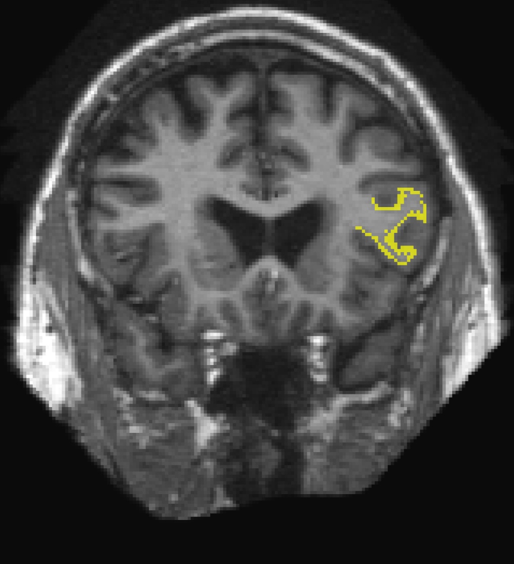
Then on the menu bar click File > Load ROI  choose lh.BA45.label and hit 'Open'. The label is visible in coronal slice 158. To jump to that slice, double click on the coordinates [127,127,128] next to where it says 'orig' in the Cursor window pane. The last number is the slice number. Change it to 158 and hit enter.

This label can be edited when the 'ROI edit'  button is chosen. Click with your left mouse button to add voxels. Hold down the shift key while using your left mouse button to delete from the label. If this was your own subject, you would want to make sure you save your edits by clicking on the 'Save ROI' button but it's not necessary for this tutorial.
To load the label on the surface, first load the the subject's inflated surface in freeview (in another terminal window) using the command below. At the top menu bar, select the 3D view.
freeview -f 004/surf/lh.inflated
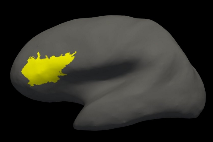
On the left menu, click on the drop down menu next to 'Curvature' and select 'Off'. Next to 'Label', select 'Load from file...'. Navigate to the subject's label directory (look one directory above) and select lh.BA45.label. Hit 'OK'. The label loaded on the inflated surface will look like this:

1.4. Individual Stats files
During the FreeSurfer processing stream, via the recon-all script, some statistical output files are generated. They are kept in each subjects' stats/ subdirectory and are generated for the subcortical segmentation (aseg) and the cortical parcellation (aparc). These tables include information on each labeled region for the individual subject.
1.4.1. aseg.stats
The statistical output from the subcortical segmentation, called aseg.stats, is a regular text file and will contain the volumes of specific structures. For example, you can obtain information such as the volume of left hippocampus and its mean intensity from this file.
cd $SUBJECTS_DIR/004/stats
less aseg.stats
At the head of the text file there will be information about the command that was run, the version used, the user who ran it and a time stamp. Following this there is information about the volume of the entire brain.
The next section of this file defines the column headers, field name, and units for the rest of the table. We can expect to see the Segmentation Id, Number of Voxels, Volume, Structure Name, Intensity normMean, Intensity normStdDev, Intensity normMin, Intensity normMax, and Intensity normRange for each entry in the table.
The remainder of the table shows this information for all the structures that are labeled in the aseg.
# Title Segmentation Statistics
#
# generating_program mri_segstats
# cvs_version $Id: mri_segstats.c,v 1.75.2.9 2013/02/16 00:09:33 greve Exp $
# cmdline mri_segstats --seg mri/aseg.mgz --sum stats/aseg.stats --pv mri/norm.mgz --empty --brainmask mri/brainmask.mgz --brain-vol-from-seg --excludeid 0 --excl-ctxgmwm --supratent --subcortgray --in mri/norm.mgz --in-intensity-name norm --in-intensity-units MR --etiv --surf-wm-vol --surf-ctx-vol --totalgray --euler --ctab /usr/local/freesurfer/stable5/ASegStatsLUT.txt --subject 004
# sysname  Linux
# hostname compute-0-2
# machine  x86_64
# user     nicks
# anatomy_type volume
#
# SUBJECTS_DIR /autofs/space/birn_045/users/BWH/buckner_data/group_study_fs5.3.0_unedited
# subjectname 004
# Measure BrainSeg, BrainSegVol, Brain Segmentation Volume, 1251739.000000, mm^3
# Measure BrainSegNotVent, BrainSegVolNotVent, Brain Segmentation Volume Without Ventricles, 1174757.000000, mm^3
# Measure BrainSegNotVentSurf, BrainSegVolNotVentSurf, Brain Segmentation Volume Without Ventricles from Surf, 1173733.938092, mm^3
# Measure lhCortex, lhCortexVol, Left hemisphere cortical gray matter volume, 251412.172031, mm^3
# Measure rhCortex, rhCortexVol, Right hemisphere cortical gray matter volume, 254032.426267, mm^3
# Measure Cortex, CortexVol, Total cortical gray matter volume, 505444.598297, mm^3
# Measure lhCorticalWhiteMatter, lhCorticalWhiteMatterVol, Left hemisphere cortical white matter volume, 241059.545562, mm^3
# Measure rhCorticalWhiteMatter, rhCorticalWhiteMatterVol, Right hemisphere cortical white matter volume, 241151.794233, mm^3
# Measure CorticalWhiteMatter, CorticalWhiteMatterVol, Total cortical white matter volume, 482211.339794, mm^3
# Measure SubCortGray, SubCortGrayVol, Subcortical gray matter volume, 67807.000000, mm^3
# Measure TotalGray, TotalGrayVol, Total gray matter volume, 665411.598297, mm^3
# Measure SupraTentorial, SupraTentorialVol, Supratentorial volume, 1128904.938092, mm^3
# Measure SupraTentorialNotVent, SupraTentorialVolNotVent, Supratentorial volume, 1057191.938092, mm^3
# Measure SupraTentorialNotVentVox, SupraTentorialVolNotVentVox, Supratentorial volume voxel count, 1056128.000000, mm^3
# Measure Mask, MaskVol, Mask Volume, 1751718.000000, mm^3
# Measure BrainSegVol-to-eTIV, BrainSegVol-to-eTIV, Ratio of BrainSegVol to eTIV, 0.697592, unitless
# Measure MaskVol-to-eTIV, MaskVol-to-eTIV, Ratio of MaskVol to eTIV, 0.976229, unitless
# Measure lhSurfaceHoles, lhSurfaceHoles, Number of defect holes in lh surfaces prior to fixing, 62, unitless
# Measure rhSurfaceHoles, rhSurfaceHoles, Number of defect holes in rh surfaces prior to fixing, 59, unitless
# Measure SurfaceHoles, SurfaceHoles, Total number of defect holes in surfaces prior to fixing, 121, unitless
# Measure EstimatedTotalIntraCranialVol, eTIV, Estimated Total Intracranial Volume, 1794371.704798, mm^3
# SegVolFile mri/aseg.mgz
# SegVolFileTimeStamp  2013/05/02 20:53:18
# ColorTable /usr/local/freesurfer/stable5/ASegStatsLUT.txt
# ColorTableTimeStamp 2013/05/03 00:10:45
# InVolFile  mri/norm.mgz
# InVolFileTimeStamp  2013/05/02 14:35:53
# InVolFrame 0
# PVVolFile  mri/norm.mgz
# PVVolFileTimeStamp  2013/05/02 14:35:53
# Excluding Cortical Gray and White Matter
# ExcludeSegId 0 2 3 41 42
# VoxelVolume_mm3 1
-
-
-
# TableCol  1 ColHeader Index
# TableCol  1 FieldName Index
# TableCol  1 Units     NA
# TableCol  2 ColHeader SegId
# TableCol  2 FieldName Segmentation Id
# TableCol  2 Units     NA
# TableCol  3 ColHeader NVoxels
# TableCol  3 FieldName Number of Voxels
# TableCol  3 Units     unitless
# TableCol  4 ColHeader Volume_mm3
# TableCol  4 FieldName Volume
# TableCol  4 Units     mm^3
# TableCol  5 ColHeader StructName
# TableCol  5 FieldName Structure Name
# TableCol  5 Units     NA
# TableCol  6 ColHeader normMean
# TableCol  6 FieldName Intensity normMean
# TableCol  6 Units     MR
# TableCol  7 ColHeader normStdDev
# TableCol  7 FieldName Itensity normStdDev
# TableCol  7 Units     MR
# TableCol  8 ColHeader normMin
# TableCol  8 FieldName Intensity normMin
# TableCol  8 Units     MR
# TableCol  9 ColHeader normMax
# TableCol  9 FieldName Intensity normMax
# TableCol  9 Units     MR
# TableCol 10 ColHeader normRange
# TableCol 10 FieldName Intensity normRange
# TableCol 10 Units     MR
# NRows 45
# NTableCols 10
-
-
-
# ColHeaders  Index SegId NVoxels Volume_mm3 StructName normMean normStdDev normMin normMax normRange
  1   4     31578    31578.0  Left-Lateral-Ventricle            12.8156    11.2321     0.0000   108.0000   108.0000
  2   5      1239     1238.5  Left-Inf-Lat-Vent                 29.4357    14.8030     0.0000    83.0000    83.0000
  3   7     13154    13154.3  Left-Cerebellum-White-Matter      84.4121     8.8272    25.0000   120.0000    95.0000
  4   8     46512    46512.4  Left-Cerebellum-Cortex            55.7117    10.1932     5.0000   116.0000   111.0000
  5  10      6438     6438.4  Left-Thalamus-Proper              83.5475    10.9499    33.0000   126.0000    93.0000
  6  11      5867     5866.8  Left-Caudate                      69.1854    12.1509    32.0000   105.0000    73.0000
  7  12      8533     8533.1  Left-Putamen                      72.5590    10.4282    11.0000   102.0000    91.0000
-
-
-
1.4.2. aparc.stats
The statistical output from the cortical parcellation, called lh.aparc.stats and rh.aparc.stats, is a regular text file and will contain the thickness of specific structures. For example, you can obtain information such as, how big is left superior temporal gyrus and its average thickness from this file.
cd $SUBJECTS_DIR/004/stats
less lh.aparc.stats
This file takes the same format as the aseg.stats. The measures at the top show the number of vertices in the cortex (NumVert) and the surface area of the cortex (SurfArea). This part of the file also tells us that the lh.aparc.annot is being used as the annotation file (AnnotationFile ../label/lh.aparc.annot).
The next section of this file defines the column headers, field name, and units for the rest of the table. We can expect to see the Structure Name, Number of Vertices, Surface Area, Gray Matter Volume, Average Thickness, Thickness StDev, Integrated Rectified Mean Curvature, Integrated Rectified Gaussian Curvature, Folding Index and Intrinsic Curvature Index for each entry in the table.
The remainder of the table shows this information for all the structures that are labeled in the aparc.
# Table of FreeSurfer cortical parcellation anatomical statistics
#
# CreationTime 2013/05/03-04:15:35-GMT
# generating_program mris_anatomical_stats
# cvs_version $Id: mris_anatomical_stats.c,v 1.72 2011/03/02 00:04:26 nicks Exp $
# mrisurf.c-cvs_version $Id: mrisurf.c,v 1.693.2.6 2013/04/26 19:03:01 nicks Exp $
# cmdline mris_anatomical_stats -mgz -cortex ../label/lh.cortex.label -f ../stats/lh.aparc.stats -b -a ../label/lh.aparc.annot -c ../label/aparc.annot.ctab 004 lh white
# sysname  Linux
# hostname compute-0-2
# machine  x86_64
# user     nicks
#
# SUBJECTS_DIR /autofs/space/birn_045/users/BWH/buckner_data/group_study_fs5.3.0_unedited
# anatomy_type surface
# subjectname 004
# hemi lh
# AnnotationFile ../label/lh.aparc.annot
# AnnotationFileTimeStamp 2013/05/03 00:02:20
# Measure Cortex, NumVert, Number of Vertices, 143074, unitless
# Measure Cortex, WhiteSurfArea, White Surface Total Area, 96267.8, mm^2
# Measure Cortex, MeanThickness, Mean Thickness, 2.29053, mm
-
-
-
# NTableCols 10
# TableCol  1 ColHeader StructName
# TableCol  1 FieldName Structure Name
# TableCol  1 Units     NA
# TableCol  2 ColHeader NumVert
# TableCol  2 FieldName Number of Vertices
# TableCol  2 Units     unitless
# TableCol  3 ColHeader SurfArea
# TableCol  3 FieldName Surface Area
# TableCol  3 Units     mm^2
# TableCol  4 ColHeader GrayVol
# TableCol  4 FieldName Gray Matter Volume
# TableCol  4 Units     mm^3
# TableCol  5 ColHeader ThickAvg
# TableCol  5 FieldName Average Thickness
# TableCol  5 Units     mm
# TableCol  6 ColHeader ThickStd
# TableCol  6 FieldName Thickness StdDev
# TableCol  6 Units     mm
# TableCol  7 ColHeader MeanCurv
# TableCol  7 FieldName Integrated Rectified Mean Curvature
# TableCol  7 Units     mm^-1
# TableCol  8 ColHeader GausCurv
# TableCol  8 FieldName Integrated Rectified Gaussian Curvature
# TableCol  8 Units     mm^-2
# TableCol  9 ColHeader  FoldInd
# TableCol  9 FieldName  Folding Index
# TableCol  9 Units      unitless
# TableCol 10 ColHeader CurvInd
# TableCol 10 FieldName Intrinsic Curvature Index
# TableCol 10 Units     unitless
-
-
-
# ColHeaders StructName NumVert SurfArea GrayVol ThickAvg ThickStd MeanCurv GausCurv FoldInd CurvInd
bankssts                                 1602   1100   2501  2.210 0.421     0.143     0.052       21     3.6
caudalanteriorcingulate                  1364    880   2416  2.404 0.712     0.172     0.085       35     4.5
caudalmiddlefrontal                      3445   2280   6543  2.514 0.505     0.155     0.083       68    10.4
cuneus                                   2573   1636   3001  1.697 0.395     0.179     0.093       61     9.8
entorhinal                                578    416   1932  3.228 0.636     0.161     0.087       11     2.3
-
-
-
1.5. Group stats files
This section will run you through using the stats directory of the subjects to perform group stats of certain structures that may be of interest to your study. The following commands will help you combine the data of the subjects you are analyzing into one table that will be easily read into a spreadsheet program. We have considered 6 subjects as examples (004, 021, 040, 067, 080, 092) in the following sections. Set your SUBJECTS_DIR to the path where you have your subjects to be analyzed.
setenv SUBJECTS_DIR $TUTORIAL_DATA/buckner_data/tutorial_subjs/group_analysis_tutorial
cd $SUBJECTS_DIR
1.5.1. Table of segmentation volumes
This section explains how to create a table of segmentation volumes using the 6 subjects mentioned above.
asegstats2table --subjects 004 021 040 067 080 092 \
  --segno 11 17 18 \
  --tablefile aseg.vol.table
where 11, 17 and 18 correspond to the segmentation label of left caudate, left hippocampus and left amygdala respectively. (You can create a table with all of the labels, not just these three, by omitting the --segno part.) Click here if you would like to view the list of labels and their corresponding ID numbers again.
The file aseg.vol.table is your output - a text file consisting of the subjects mentioned in the command above and the values for the structures requested along with the measures in the header (i.e. BrainSegVol). The information in this text file is formatted so it can be easily imported into a spreadsheet program (often used as input for many statistical analysis programs). If you do the ls command, you should see that the text file aseg.vol.table has been created. To see what the file looks like, do:
less aseg.vol.table
To load the resulting table into a spreadsheet, run:
gnumeric aseg.vol.table
Note: the gnumeric command is meant to be run on a Linux machine. Mac users could install OpenOffice 3.0 to open the table in a spreadsheet and run the command.
/Applications/OpenOffice.org.app/Contents/MacOS/scalc aseg.vol.table
In the table, the first cell is volume indicating that the measure is a volume in mm3. About the subject IDs, you'll notice that in the examples we've considered here, each subject is a 3 digit number. Therefore gnumeric thinks it is a number and removes leading 0s. This is a gnumeric issue but probably it would not happen if subject names had characters in them instead of integers.
1.5.2. Table of segmentation mean intensities
The purpose of this section is to demonstrate how you can change what measure you collect for your spreadsheet from volume to mean intensity using the asegstats2table command.
asegstats2table \
  --subjects 004 021 040 067 080 092 \
  --segno 11 17 18 \
  --meas mean \
  --tablefile aseg.mean-intensity.table
You can load the table into a spreadsheet as explained in the previous section or view aseg.mean-intensity.table with a text editor, like gedit.
1.5.3. Table of white matter parcellation volumes
The purpose of this section is to show how you can change which segmentation atlas you get stats from (and thus which structures):
asegstats2table \
  --subjects 004 021 040 067 080 092 \
  --segno 3007 3021 3022 4022 \
  --stats wmparc.stats \
  --tablefile wmparc.vol.table
This prints out stats on the white matter parcellation.
1.5.4. Table of the surface area of each cortical parcellation in the Desikan atlas
This section demonstrates how to create a table of the surface area of each cortical parcellation in the Desikan atlas (surface area is the default measure).
aparcstats2table --hemi lh \
  --subjects 004 021 040 067 080 092 \
  --tablefile lh.aparc.area.table
Feel free to take a look at those results.
1.5.5. Table of the average thickness of each cortical parcellation in the Destrieux atlas
The purpose of this section is to show how to change the summary measure (in this case, to thickness) and the parcellation atlas (to Destrieux's atlas).
aparcstats2table --hemi lh \
  --subjects 004 021 040 067 080 092 \
  --meas thickness \
  --parc aparc.a2009s \
  --tablefile lh.aparc.a2009.thickness.table
You can now load the resulting table into a spreadsheet.
