mri_3d_photo_recon -h
--input_photo_dir |
Directory with input pixel-corrected photos. |
--input_segmentation_dir |
Directory with input slab masks/segmentations |
--ref_mask |
When using a binary volume as a reference. |
--ref_surface |
Using a 3D surface scan as a reference. |
--ref_soft_mask |
Using the provided average atlas. Best for using retrospective processing on data without a better reference. |
--mesh_reorient_with_indices |
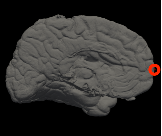
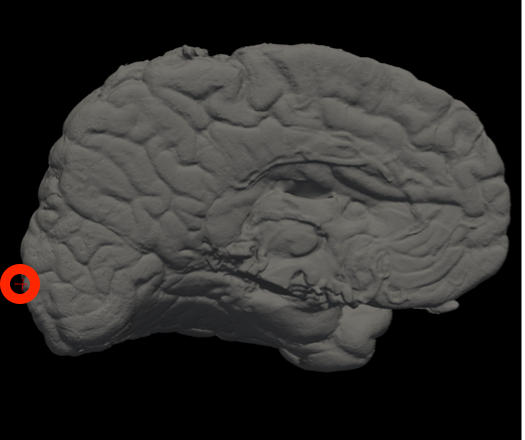
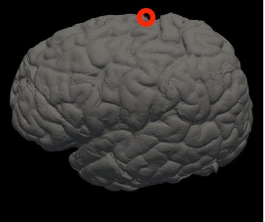
Vertex indices of the frontal pole, occipital pole, and top of the central sulcus, separated with commas, for mesh alignment. |
--photos_of_posterior_side |
Use when photos are taken of the posterior side of slabs (default is anterior side). |
--order_posterior_to_anterior |
Use when photos are ordered from posterior to anterior (default is anterior to posterior). |
--allow_z_stretch |
Use to adjust the slice thickness to best match the reference. You should probably *never* use this with soft references (ref_soft_mask). |
--rigid_only_for_photos |
Switch on if you want photos to deform only rigidly (not affine). |
--slice_thickness |
Slice thickness in mm. |
--photo_resolution |
Resolution of the photos in mm. |
||--output_directory ||Output directory with reconstructed photo volume and reference ||
In Freeview, you can find the number corresponding to the vertices of each anatomical area to use in the --mesh_reorient_with_indices flag. These indices should be comma-separated in the order: frontal pole, occipital pole, and top of the central sulcus.
|
|
|
Frontal Pole |
Occipital Pole |
Top of the central sulcus |
For example, the --mesh_reorient_with_indices flag might look like this:
- –mesh_reorient_with_indices 999999,999999,999999
Example for running reconstruction
- mri_3d_photo_recon \
- --input_photo_dir ./deformed \
- --input_segmentation_dir ./connected_components \
- --ref_surface ./mesh/case.stl \
- --mesh_reorient_with_indices 999,1010,333333 \
- --photos_of_posterior_side --allow_z_stretch \
- --slice_thickness 8 \
- --photo_resolution 0.1


