| Deletions are marked like this. | Additions are marked like this. |
| Line 2: | Line 2: |
|
''Authors: Henry Tregidgo and Juan Eugenio Iglesias'' <<BR>> ''E-mail: h.tregidgo [at] ucl.ac.uk and jiglesiasgonzalez [at] mgh.harvard.edu'' <<BR>> <<BR>> ''Rather than directly contacting the authors, please post your questions on this module to the FreeSurfer mailing list at freesurfer [at] nmr.mgh.harvard.edu'' <<BR>> <<BR>> If you use these tools in your analysis, please cite: * [[https://www.sciencedirect.com/science/article/pii/S1053811923002744|Accurate Bayesian segmentation of thalamic nuclei using diffusion MRI and an improved histological atlas]]. Tregidgo HFJ, Soskic S, Althonayan J, Maffei C, Van Leemput K, Golland P, Insausti R, Lerma-Usabiaga G, Caballero-Gaudes C, Paz-Alonso PM, Yendiki A, Alexander DC, Bocchetta M, Rohrer JD, Iglesias JE. Neuroimage (accepted). |
'''''We are working on integrating the Bayesian implementation of this method into the Python framework that unifies the subregion segmentation modules.''''' ''Authors: Henry Tregidgo and Juan Eugenio Iglesias'' <<BR>> ''E-mail: h.tregidgo [at] ucl.ac.uk and jiglesiasgonzalez [at] mgh.harvard.edu'' <<BR>> <<BR>> ''Rather than directly contacting the author, please post your questions on this module to the FreeSurfer mailing list at freesurfer [at] nmr.mgh.harvard.edu'' <<BR>> <<BR>> If you use these tools in your analysis, please cite: * [[https://doi.org/10.1016/j.neuroimage.2023.120129|Accurate Bayesian segmentation of thalamic nuclei using diffusion MRI and an improved histological atlas]]. Tregidgo HFJ, Soskic S, Althonayan J, Maffei C, Van Leemput K, Golland P, Insausti R, Lerma-Usabiaga G, Caballero-Gaudes C, Paz-Alonso PM, Yendiki A, Alexander DC, Bocchetta M, Rohrer JD, Iglesias JE., NeuroImage, 274, 120129,(2023) Additionally for the CNN implementation: * [[https://arxiv.org/|Domain-agnostic segmentation of thalamic nuclei from joint structural and diffusion MRI]]. Tregidgo HFJ, Soskic S, Olchany MD, Althonayan J, Billot B, Maffei C, Golland P, Yendiki A, Alexander DC, Bocchetta M, Rohrer JD, Iglesias JE. (Under review) |
| Line 6: | Line 24: |
|
<<BR>> === Contents === 1. Motivation and General Description 2. Installation 3. Usage 4. Gathering the volumes from all analyzed subjects 5. Frequently asked questions 6. Multimodal integration 7. Test data <<BR>> === 1. Motivation and General Description === The [[https://freesurfer.net/fswiki/ThalamicNuclei|ThalamicNuclei subfields module]] produces a parcellation of the thalamus into 25 different nuclei by applying a probabilistic atlas built with histological data to structural MRI volumes. However the lack of contrast in some structural scans can cause difficulty in correctly identifying boundaries that are more easily identifiable in modalities such as diffusion weighted MRI. <<BR>> {{attachment:MissingStructuralBoundaries.png||height="300"}} <<BR>><<BR>> For this reason we have improved our method by incorporating information from diffusion imaging, specifically diffusion tensor imaging (DTI). To do this we not only incorporate a DTI likelihood term into our Bayesian framework but also modify our atlas to allow modelling of DTI specific inhomogeneities such as with white matter tracts or between the medial and lateral portions of the PuM. This results in improved identification of both interior and exterior thalamic boundaries. <<BR>> {{attachment:HCPcomparison3.png||height="400"}} <<BR>><<BR>> Additionally we address partial volume effects, present in DTI volumes with large voxels, by providing a CNN segmentation implementation. This CNN can segment the thalamic nuclei from T1 and diffusion MRI data obtained with virtually any acquisition, solving the problems posed by PV effects to Bayesian segmentations. <<BR>> {{attachment:examples.pdf||height="270"}} <<BR>><<BR>> === 2. Bayesian Tool Usage === This module is currently being integrated into the Python framework that unifies the subregion segmentation modules and will be available soon. <<BR>> === 3. CNN Tool Usage === This software requires four registered input volumes. These are: 1. A bias corrected whole brain T1 such as the norm.mgz output from the main !FreeSurfer stream ("recon-all") 2. A coarse whole brain segmentation such as aseg.mgz 3. A fractional anisotropy volume such as dtifit_FA.nii.gz produced by the [[https://surfer.nmr.mgh.harvard.edu/fswiki/Tracula|TRACULA]] module 4. A 4D volume containing the principle direction vector for each DTI voxel (eg. dtifit_V1.nii.gz from [[https://surfer.nmr.mgh.harvard.edu/fswiki/Tracula|TRACULA]]) |
|
| Line 8: | Line 68: |
| This module is coming soon... |
An example workflow for obtaining these volumes would be: {{{ recon-all -all -s <subjid> }}} where <subjid> is the name of the subject. {{{ trac-all -prep -s <subjid> -I <dicomfile> }}} where <dicomfile> is the path to the diffusion weighted MRI dicoms (see [[https://surfer.nmr.mgh.harvard.edu/fswiki/trac-all#Usage|trac-all]]). {{{ mri_convert <subjid>/mri/norm.mgz <subjid>/mri/norm.aligned.mgz -at <subjid>/dmri/xfms/anatorig2diff.bbr.lta mri_convert <subjid>/mri/aseg.mgz <subjid>/mri/aseg.aligned.mgz -at <subjid>/dmri/xfms/anatorig2diff.bbr.lta }}} {{{ mri_segmentThalamicNucleiDTI_CNN --t1 <subjid>/mri/norm.aligned.mgz --aseg <subjid>/mri/aseg.aligned.mgz --fa <subjid>/dmri/dtifit_FA.nii.gz --v1 <subjid>/dmri/dtifit_V1.nii.gz --o path/to/outputSegmentation.nii.gz --vol path/to/measuredVolumes.csv --threads <nthreads> }}} Where <nthreads> is the number of threads to use in processing. |
Joint segmentation of thalamic nuclei from T1 scan and DTI
We are working on integrating the Bayesian implementation of this method into the Python framework that unifies the subregion segmentation modules.
Authors: Henry Tregidgo and Juan Eugenio Iglesias
E-mail: h.tregidgo [at] ucl.ac.uk and jiglesiasgonzalez [at] mgh.harvard.edu
Rather than directly contacting the author, please post your questions on this module to the FreeSurfer mailing list at freesurfer [at] nmr.mgh.harvard.edu
If you use these tools in your analysis, please cite:
* Accurate Bayesian segmentation of thalamic nuclei using diffusion MRI and an improved histological atlas. Tregidgo HFJ, Soskic S, Althonayan J, Maffei C, Van Leemput K, Golland P, Insausti R, Lerma-Usabiaga G, Caballero-Gaudes C, Paz-Alonso PM, Yendiki A, Alexander DC, Bocchetta M, Rohrer JD, Iglesias JE., NeuroImage, 274, 120129,(2023)
Additionally for the CNN implementation:
* Domain-agnostic segmentation of thalamic nuclei from joint structural and diffusion MRI. Tregidgo HFJ, Soskic S, Olchany MD, Althonayan J, Billot B, Maffei C, Golland P, Yendiki A, Alexander DC, Bocchetta M, Rohrer JD, Iglesias JE. (Under review)
See also: ThalamicNuclei
Contents
1. Motivation and General Description 2. Installation 3. Usage 4. Gathering the volumes from all analyzed subjects 5. Frequently asked questions 6. Multimodal integration 7. Test data
1. Motivation and General Description
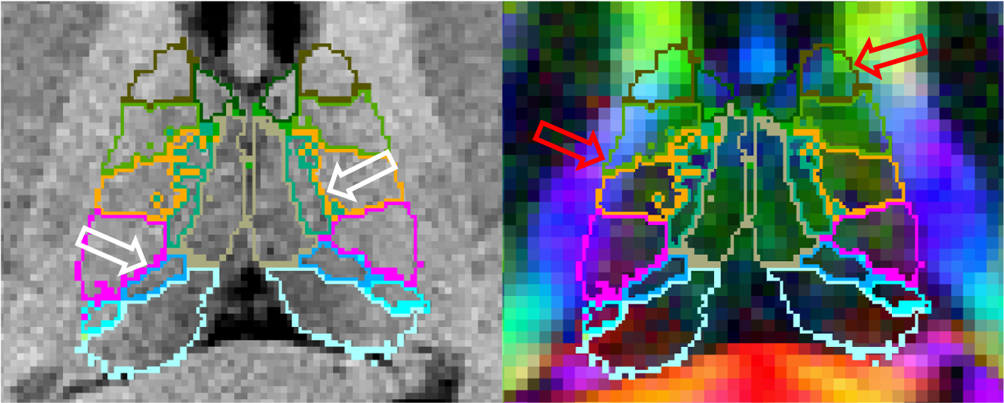
The ThalamicNuclei subfields module produces a parcellation of the thalamus into 25 different nuclei by applying a probabilistic atlas built with histological data to structural MRI volumes. However the lack of contrast in some structural scans can cause difficulty in correctly identifying boundaries that are more easily identifiable in modalities such as diffusion weighted MRI.

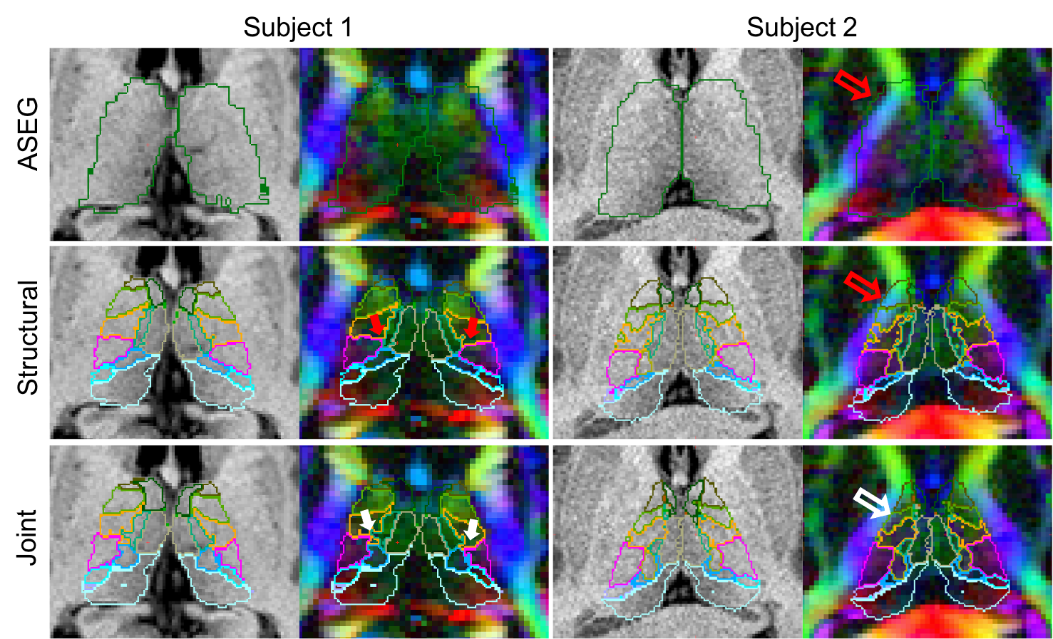
For this reason we have improved our method by incorporating information from diffusion imaging, specifically diffusion tensor imaging (DTI). To do this we not only incorporate a DTI likelihood term into our Bayesian framework but also modify our atlas to allow modelling of DTI specific inhomogeneities such as with white matter tracts or between the medial and lateral portions of the PuM. This results in improved identification of both interior and exterior thalamic boundaries.

Additionally we address partial volume effects, present in DTI volumes with large voxels, by providing a CNN segmentation implementation. This CNN can segment the thalamic nuclei from T1 and diffusion MRI data obtained with virtually any acquisition, solving the problems posed by PV effects to Bayesian segmentations.
2. Bayesian Tool Usage
This module is currently being integrated into the Python framework that unifies the subregion segmentation modules and will be available soon.
3. CNN Tool Usage
This software requires four registered input volumes. These are: 1. A bias corrected whole brain T1 such as the norm.mgz output from the main FreeSurfer stream ("recon-all") 2. A coarse whole brain segmentation such as aseg.mgz 3. A fractional anisotropy volume such as dtifit_FA.nii.gz produced by the TRACULA module 4. A 4D volume containing the principle direction vector for each DTI voxel (eg. dtifit_V1.nii.gz from TRACULA)
An example workflow for obtaining these volumes would be:
recon-all -all -s <subjid>
where <subjid> is the name of the subject.
trac-all -prep -s <subjid> -I <dicomfile>
where <dicomfile> is the path to the diffusion weighted MRI dicoms (see trac-all).
mri_convert <subjid>/mri/norm.mgz <subjid>/mri/norm.aligned.mgz -at <subjid>/dmri/xfms/anatorig2diff.bbr.lta
mri_convert <subjid>/mri/aseg.mgz <subjid>/mri/aseg.aligned.mgz -at <subjid>/dmri/xfms/anatorig2diff.bbr.lta
mri_segmentThalamicNucleiDTI_CNN --t1 <subjid>/mri/norm.aligned.mgz --aseg <subjid>/mri/aseg.aligned.mgz --fa <subjid>/dmri/dtifit_FA.nii.gz --v1 <subjid>/dmri/dtifit_V1.nii.gz --o path/to/outputSegmentation.nii.gz --vol path/to/measuredVolumes.csv --threads <nthreads>
Where <nthreads> is the number of threads to use in processing.
